Brain tumors present a complex and challenging medical condition that requires precise diagnosis and targeted treatment. Over the years, significant advancements have been made in brain tumor imaging techniques, revolutionizing how these tumors are diagnosed and monitored. Through innovative imaging modalities, healthcare professionals can obtain detailed information about brain tumor location, size, and characteristics, leading to more accurate diagnoses and personalized treatment plans.
This article will explore the latest advancements in brain tumor imaging and their impact on improving patient outcomes.
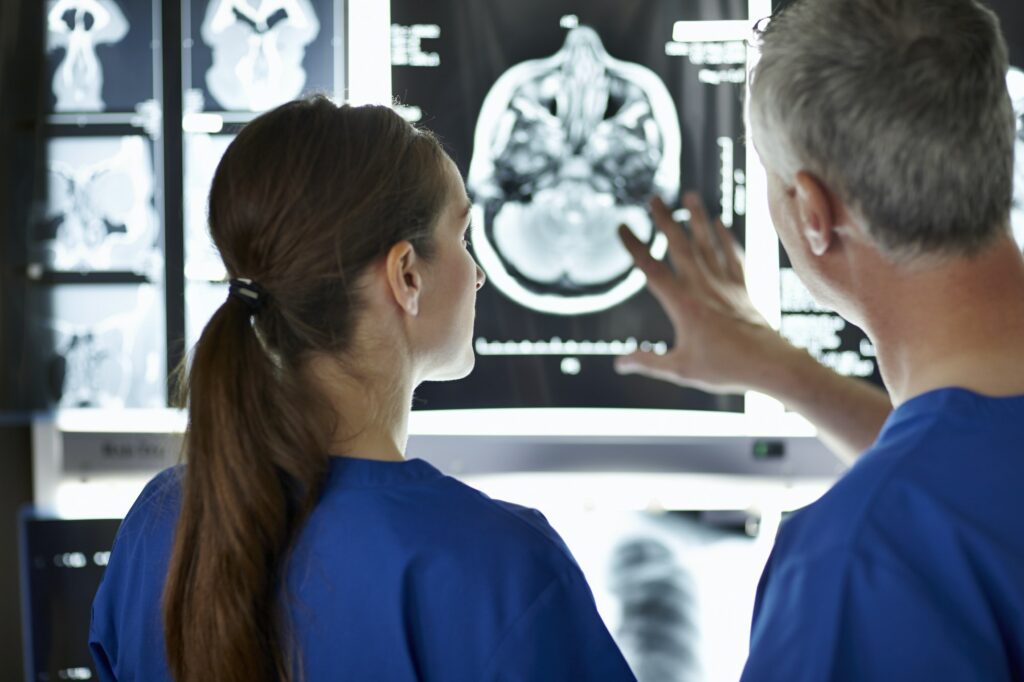
Magnetic Resonance Imaging (MRI):
Magnetic Resonance Imaging (MRI) has emerged as a cornerstone in brain tumor diagnosis and assessment. This non-invasive imaging technique utilizes powerful magnets and radio waves to generate detailed brain images. MRI provides high-resolution images that allow healthcare professionals to visualize brain tumors and assess their characteristics, such as size, location, and involvement of surrounding structures. The development of advanced MRI sequences, such as diffusion-weighted imaging (DWI) and perfusion imaging, has further enhanced the accuracy of tumor detection and characterization.
For brain tumors, MRI is often used to:
- Diagnose the tumor: MRI can often be used to diagnose a brain tumor, even if it is small or located in a difficult-to-see area.
- Determine the size and location of the tumor: MRI can be used to determine the size and location of a brain tumor, which is important for planning treatment.
- Assess the tumor’s characteristics: MRI can be used to assess the characteristics of a brain tumor, such as its type, grade, and whether it has spread to other parts of the brain.
- Guide biopsy or surgery: MRI can be used to guide biopsy or surgery, which are two types of treatments for brain tumors
Functional MRI (fMRI):
Functional MRI (fMRI) is a specialized imaging technique that provides insights into the brain’s functional areas, allowing healthcare professionals to map critical regions affected by tumors. By measuring blood flow changes, fMRI can identify areas involved in essential functions such as speech, movement, and memory. This information is crucial in surgical planning, as it helps surgeons navigate around critical regions and minimize the risk of functional deficits during tumor removal.
Positron Emission Tomography (PET):
Positron Emission Tomography (PET) imaging is vital in assessing brain tumor metabolism and identifying areas of increased cellular activity. PET scans involve the injection of a radioactive tracer that targets glucose or other specific molecules within the tumor. The tracer emits positrons, which the PET scanner detects to create a three-dimensional image of tumor activity. PET scans can help differentiate between benign and malignant tumors, assess tumor grade and aggressiveness, and monitor treatment response.
Single-Photon Emission Computed Tomography (SPECT):
Single-Photon Emission Computed Tomography (SPECT) is another valuable imaging modality used in brain tumor evaluation. SPECT scans involve the injection of a radioactive tracer that emits gamma rays, which are detected by a specialized camera. By analyzing the distribution of the tracer, SPECT provides information about blood flow, metabolism, and receptor activity within the brain. SPECT imaging can assist in tumor localization, distinguishing between tumor recurrence and radiation necrosis, and evaluating treatment response.
SPECT can be used to assess the following aspects of brain tumors:
- Tumor location: SPECT can be used to identify the location of a brain tumor, even if it is small or located in a difficult-to-see area.
- Tumor activity: SPECT can be used to assess the activity of a brain tumor, which can help to determine whether it is growing or shrinking.
- Tumor recurrence: SPECT can be used to distinguish between tumor recurrence and radiation necrosis, which is a condition that can occur after radiation therapy.
- Treatment response: SPECT can be used to evaluate the response of a brain tumor to treatment.
Advanced Imaging Techniques:
In addition to the imaging mentioned above modalities, several advanced techniques have emerged to improve the accuracy and specificity of brain tumor diagnosis. For instance, spectroscopy is a technique that measures the chemical composition of tissues, helping to differentiate between tumor types and assess tumor boundaries. Diffusion Tensor Imaging (DTI) allows visualization of nerve fiber pathways, aiding surgeons in planning tumor resection while preserving critical neural connections. Perfusion-weighted imaging provides information about blood flow within tumors, helping to evaluate tumor grade and monitor treatment response.
Radiomics and Artificial Intelligence (AI):
Radiomics and Artificial Intelligence (AI) have the potential to revolutionize brain tumor imaging analysis. Radiomics involves extracting and analyzing a vast array of quantitative imaging features from medical images. These features provide valuable information about tumor characteristics, heterogeneity, and treatment response. AI algorithms can then analyze this data to aid diagnosis, treatment planning, and predictive assessment. AI-powered tools also can integrate imaging data with genomic information, facilitating precision medicine approaches for individualized treatment strategies.

Radiomics and AI are still in their early stages of development, but they have the potential to revolutionize brain tumor imaging analysis. These technologies have the potential to improve diagnosis, treatment planning, and predictive assessment for patients with brain tumors.
Here are some of the benefits of using radiomics and AI in brain tumor imaging:
- Improved accuracy of diagnosis: Radiomics and AI can be used to identify tumors that are difficult to see on traditional imaging scans. This can lead to earlier diagnosis and treatment, which can improve patient outcomes.
- Better understanding of tumor biology: Radiomics and AI can be used to identify the molecular characteristics of tumors. This information can be used to develop more targeted and effective treatments.
- Personalized treatment planning: Radiomics and AI can be used to predict how a patient will respond to different treatments. This information can be used to personalize treatment plans and improve patient outcomes.
Conclusion:
Advancements in brain tumor imaging techniques have transformed how healthcare professionals diagnose, characterize, and treat these complex tumors. MRI, fMRI, PET, SPECT, advanced imaging techniques, and the integration of radionics and AI have collectively revolutionized the field. These innovations enable more precise diagnosis, improved surgical planning, better treatment monitoring, and the development of personalized therapeutic strategies. As technology advances, brain tumor imaging will continue to evolve, further enhancing patient care and outcomes.